


A sliver of lung tissue glows faintly under a microscope, and within minutes an algorithm reads the pattern of that light and predicts whether a patient’s cancer carries a mutation that changes how it should be treated. That is the promise behind a method developed by scientists at the University of Edinburgh and NHS Lothian, and if it holds up in wider testing, it could reshape how quickly and cheaply lung cancer gets diagnosed.
Reading light instead of sequencing genes
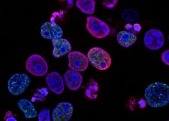
The technique relies on fluorescence lifetime imaging microscopy, known as FLIM, which captures the natural light signals given off by tissue. Rather than sending a biopsy through the slow, costly process of genetic sequencing, the researchers trained an AI model to spot patterns in those light signals that correspond to specific DNA changes. In the study, the method predicted mutations in the EGFR gene, a key early indicator of lung cancer, and went a step further by telling apart the two most common EGFR mutation types that clinicians use to decide on treatment.
That second part is what makes it clinically interesting. Knowing whether a mutation is present is useful. Knowing which mutation it is can point a patient toward one targeted therapy over another, and getting that answer in minutes rather than weeks changes what a clinic can realistically offer.
The cost problem it targets
Molecular testing is expensive, it is slow, and it eats through something in short supply: tissue. Biopsy samples tend to be tiny, and every test carves into a limited amount of material. Dr Qiang Wang, co-lead of the study from Edinburgh’s Institute for Regeneration and Repair, framed the potential savings in plain terms. “This approach has the potential to take processes that currently cost thousands of pounds and require weeks of lab work and reduce them to something that takes minutes and costs hundreds,” he said. “That is a step change in what is clinically achievable, particularly for centres and health systems where access to complex molecular testing is limited.”
Because FLIM does not damage the tissue it reads, the biopsy stays intact and available for further analysis. For pathologists working with fragments no larger than a grain of rice, keeping the sample whole while still extracting an answer is close to having it both ways.
Pressure on diagnostic services
The timing speaks to a wider strain. Dr David Dorward, a consultant thoracic pathologist at NHS Lothian, said the volume of work is climbing. “Clinicians are increasingly seeing more patients with earlier-stage disease and dealing with a growing number of biopsy samples, placing significant pressure on diagnostic services,” he said. “Technologies like this, which can deliver more information from smaller tissue samples at speed, will be essential for developing clinically effective diagnostic pathways.”
The stakes in Scotland alone are heavy. According to the most recent figures from Public Health Scotland, lung cancer remained the most common cause of cancer death in 2024, with more than 3,650 deaths, roughly a fifth of all cancer deaths in the country. Most of those, the agency notes, could be avoided by eliminating smoking. Faster, cheaper detection does not change that underlying cause, but it can widen the window in which treatment still works.
A pattern of AI moving into Scottish hospitals
This is not the first time an AI tool has shown its worth inside NHS Scotland. A breast screening system called GEMINI, run by NHS Grampian and the University of Aberdeen, analysed more than 10,000 mammograms in 2023. Research found it lifted breast cancer detection rates by more than 10 percentage points and cut the time to notify affected women from 14 days to just three. Results like that explain why hospitals keep saying yes to these pilots.
The enthusiasm comes with a caution attached. The innovation agency InnoScot Health warned earlier this year that folding AI into the NHS is a significant challenge for decision-makers, even with the huge promise the technology carries. Their point is that speed and accuracy are only half the equation. The other half is governance, and whether patients trust a system that lets software weigh in on a cancer diagnosis.
The next test is scale. A method that works on a research cohort still has to prove itself across thousands of real patients, different scanners, and the messy variety of everyday clinical samples before it earns a permanent place in the pathway. If it clears that bar, the more interesting question is which other cancers a light-reading algorithm could learn to spot. For more coverage of AI in healthcare, visit Mylistingo.
The Netherlands, for internationals
One email a week: the week's news for internationals in the Netherlands, new practical guides and what changed in the cities. No spam, unsubscribe any time.